The service of Gastroenterology at the National Center for Minimally Invasive Surgery has over 15 years of experience in endoscopic procedures. This team of professors and specialists trained at the Cuban school of gastroenterology and digestive endoscopy, has also been acknowledged though courses and training in several worldwide high level centers in the specialty, such as: North Tyneside General Hospital of Newcastle, England; Jichi Medical School, Toho University Omori Hospital and National Cancer Center in Japan; Clínica Quirón and Universidad Autónoma de Barcelona, Tuebingen University in Germany, Hospital Clinic and St Creu et Sau Pau and Val de Hebron de Barcelona, Spain and Hospital das Clinicas, School of Medicine, University of Sao Paulo, Brazil. This team has performed a total of 39 426 studies with different endoscopic techniques, and it is a pioneer in the introduction of new diagnostic and therapeutic techniques in the endoscopy of the digestive system in Cuba.
Endoscopic Ampulectomy | ERCP - Pancreatic Lithiasis Extraction | ERCP - Lithiasis Extraction in Bile Duct | Pseudocyst drainage |
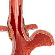
Esophageal stenosis is a narrowing in any portion of the esophagus that causes difficulty or
obstruction to the passing of food towards the gastric cavity, defined as dysphagia.
Due to its cause, it may be defined as:
Benign: peptic esophagitis, caustic esophagitis, esophagitis brought on by medication, infectious esophagitis and iatrogenic esophagitis. It usually starts slowly or unpredictably, with antecedents of diseases or prior accidents.
Malignant: Obstruction due to growth of esophageal intraluminal neoplastic lesions; they can be concentric, exophytic, or coming from the muscle wall or submucosa; due to compression or invasion of the mediastinal process; gradual appearance, progressive, first to solids, later to fluids until its complete occlusion, accompanied by loss of weight and other general symptoms.
Physician Team:
Julián Francisco Ruiz Torres, MD, PhD
Jorge L. García Menocal, MD
Contact: tursalud@cce.sld.cu
The ingestion of foreign bodies most frequently occur in the upper digestive tract (esophagus,
stomach or duodenum, in that order), but can also be seen in the small intestine, colon or rectum
The symptoms will depend on the nature of the object, its location, entry pathway, and time elapsed
after it is inside the organism. It is estimated that around 80% of the total number of ingested
foreign bodies are spontaneously eliminated through the feces because they pass along the digestive
tract without difficulty. According to the different series that have been published, extraction by
means of an endoscope is performed in around 19% of the cases, and only 1% would require surgical
extraction.
Usually, they are food particles. Meat is the most common responsible agent, whether because it is a
chunk which is too large to pass along a normal esophagus, or because, even if it is small, it has
been stopped by a pathological obstacle, which could be stenosis, a tumor, an angled or L-shaped
esophagus.
Upper digestive tract (UDT): Esophageal, gastric or duodenal
Middle digestive tract (MDT): Small intestine, posterior to 2nd duodenal portion down
to the ileocecal valve
Lower digestive tract (LDT): CColon and rectum
Physician Team:
Julián Francisco Ruiz Torres, MD, PhD
Vivianne Anido Escobar, MD, PhD
Jorge L. García Menocal, MD
Felipe Neri Piñol Jiménez, MD, PhD
Norberto Alfonso Contino, MD, PhD
Mildred Cecilia Armenteros, MD
Contact to: tursalud@cce.sld.cu
Digestive hemorrhage is defined as the blood loss coming from the digestive system. It is one of the
most frequent medical emergencies being responsible for a large number of hospitalizations, with a
mortality ranging between 5% and 20%, which varies depending on different factors, especially the
amount of bleeding, its origin, age of the patient and other associated diseases.
Digestive hemorrhage, depending on its origin above or below Treitz arch, is classified as upper
digestive (UDH) or lower digestive hemorrhage (LDH).
Specific treatment of the bleeding lesion is directed towards inhibiting active hemorrhage and
avoiding relapse. It is divided based on its origin as variceal if it depends on esophageal or gastric
varices and non variceal if it is caused by the rest of the lesions in this path.
Physician Team:
Julián Francisco Ruiz Torres, MD, PhD
Vivianne Anido Escobar, MD, PhD
Jorge L. García Menocal, MD
Felipe Neri Piñol Jiménez, MD, PhD
Norberto Alfonso Contino, MD, PhD
Mildred Cecilia Armenteros, MD
Contact to: tursalud@cce.sld.cu
Premature benign and malignant lesions of the upper digestive tract are not infrequent in daily
medical practice. During an endoscopy it is very common to see this kind of lesions; or a patient
arrives with a clinical picture that makes us suspect they are present; or he is brought from another
hospital to complete the study of a particular lesion or perform endoscopic treatment.
These lesions can appear morphologically by endoscopy in the following way:
Inflammatory lesions, polypoid lesions, raised lesions, diverticula, intraluminal tumors and extrinsic
compressions of neighboring organs.
Its clinical manifestation varies from an asymptomatic picture to episodes of bleeding and
obstructions.
For their best approach, they are divided based on their location in the upper digestive tract and on
its morphologic characteristic in benign or malignant.
Physician Team:
Julián Francisco Ruiz Torres, MD, PhD
Vivianne Anido Escobar, MD, PhD
Jorge L. García Menocal, MD
Felipe Neri Piñol Jiménez, MD, PhD
Norberto Alfonso Contino, MD, PhD
Mildred Cecilia Armenteros, MD
Contact to: tursalud@cce.sld.cu
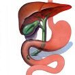
Non-lithiasic benign lesions of the common bile duct can be mostly caused during the course of diverse hepatobiliary and gastroduodenal (iatrogenic) intra-abdominal surgeries. Its incidence has increased up to 0.4% in the last decade after the introduction of laparoscopic cholecystectomy. Nevertheless, other lesions capable of obstructing the biliary flow to the duodenum have been identified such as foreign bodies (parasites), external, post-inflammatory and congenital compressions. Emphasis has been made on the different technical aspects of its treatment.
Patients who have been diagnosed or are suspicious of having non-lithiasic benign lesions of the bile ducts, such as post-surgical bile duct lesions according to Bismuth and Ballesta, biliary fistulas, foreign bodies (parasites), extrinsic compressions, biliary, pancreatic and congenital post-inflammatory stenosis, have been studied and treated for jaundice.
Physician Team:
Julián Francisco Ruiz Torres, MD, PhD
Jorge L. García Menocal, MD, PhD
Norberto Alfonso Contino, MD
Contact to: tursalud@cce.sld.cu
Physician Team:
Julián Francisco Ruiz Torres, MD, PhD
Jorge L. García Menocal, MD, PhD
Norberto Alfonso Contino, MD
Contact to: tursalud@cce.sld.cu
Patients with non-resectable extra-hepatic bile duct cancer have a cancer that cannot be completely
removed by the surgeon. These patients represent most patients with bile duct cancer. Frequently, this
cancer directly invades the portal vein, the liver or expands along the common bile ducts to the
adjacent lymph nodes. It is not common for this cancer to expand to other body areas, but
intra-abdominal metastases, mainly peritoneal metastases, occur.
At this stage, patient management is focused on palliation.
Physician Team:
Julián Francisco Ruiz Torres, MD, PhD
Jorge L. García Menocal, MD, PhD
Norberto Alfonso Contino, MD
Contact to: tursalud@cce.sld.cu
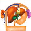
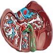
Acute Pancreatitis.
Biliary acute pancreatitis is an acute inflammatory reversible process of the pancreas caused by the obstruction of the pancreatic flow due to the presence or transit of lithiasis through the ampulla orifice coming from the biliary tree. Endoscopic retrograde cholangiopancreatography allows etiological treatment with a change in the patient ´s recovery. It is a procedure of relative emergency. At this stage, patient treatment is focused on palliation.
Chronic pancreatitis.
Chronic pancreatitis is an irreversible chronic inflammatory process of the pancreas that causes the
formation of fibrosis and the destruction of tissues with exocrine and endocrine functions.
Endoscopic retrograde cholangiopancreatography allows evaluating the status of the bile and pancreatic
ducts with high sensibility and specificity.
Physician Team:
Julián Francisco Ruiz Torres, MD, PhD
Jorge L. García Menocal, MD, PhD
Norberto Alfonso Contino, MD
Contact to: tursalud@cce.sld.cu